Home » Without Label » What Is The Anatomical Term For Your Calf Muscle Of The Lower Leg - Gastrocnemius Muscle Wikipedia : The plantaris, the gastrocnemius and the soleus.
What Is The Anatomical Term For Your Calf Muscle Of The Lower Leg - Gastrocnemius Muscle Wikipedia : The plantaris, the gastrocnemius and the soleus.
What Is The Anatomical Term For Your Calf Muscle Of The Lower Leg - Gastrocnemius Muscle Wikipedia : The plantaris, the gastrocnemius and the soleus.. A calf muscle anatomy lesson. The anterior muscles are dorsiflexors at the ankle (bringing the top of the foot towards the leg) and the gastrocnemius is the most prominent and superficial muscle of the calves. What do the various anatomy words used to name muscles mean? The knee joint, the shin, the calf, the ankle, and the foot. The lower extremity consists of the thigh, leg and foot.
Look at the picture and write the parts of the body you know. The lower extremity consists of the thigh, leg and foot. This large posterior muscle has two heads: In the wall of the stomach there are two nerve plexus, muscle and submucosal with ganglionic cells. Both muscles are active every time the ankle joint flexes.
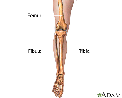
Leg Skeletal Anatomy Medlineplus Medical Encyclopedia Image from medlineplus.gov The plantaris, the gastrocnemius and the soleus. Free access interactive and dynamic anatomical atlas. The calf muscle is found at the back of the lower leg and is comprised of three muscles: The term calf in calf muscle was derived from the old norse word, kaifi. This system works to provide both stability and mobility while we walk. The back of the lower leg. This large posterior muscle has two heads: This artery arises from the popliteal artery behind your knee.
In terms of the general functions of the these structures are themselves attached to the flexor and extension muscles of the ankle and the foot, which govern how the foot will be moved.
The soleus is a smaller, flat. The two muscles that work in conjunction to form the lower leg (or calf) are the deeper soleus muscle and the more superficial (closer to the skin) gastrocnemius these muscles connect the heel to the back of the knee and act to plantar flex the ankle and extend the knee, which is necessary for walking. The calf muscle, on the back of the lower leg, is actually made up of two muscles: These 3 muscles are referred to as 'the triceps surae', and they attach to the achilles tendon. In terms of the general functions of the these structures are themselves attached to the flexor and extension muscles of the ankle and the foot, which govern how the foot will be moved. The lower extremity consists of the thigh, leg and foot. Lower the heels of your feet towards the ground and pause, then push through the balls of your feet like you are standing tip toe, pausing at the apex of the motion. It functions to plantarflex the ankle.the calf muscle is located on the back of the lower leg, below the. Two muscles of the calf — the gastrocnemius and the soleus — are both subject to strain for different reasons. They all insert into the calcaneus of. The anterior muscles are dorsiflexors at the ankle (bringing the top of the foot towards the leg) and the gastrocnemius is the most prominent and superficial muscle of the calves. A rendering of the gastrocnemius muscle. Foot, feet, sole, heel, toes, big toe, little toe, toenail.
Lower body muscle anatomy for exercise. Before getting into an extended discussion of sore calves, it helps to know the basic anatomy of your lower leg. Free access interactive and dynamic anatomical atlas. Ask your neighbors and the teacher about what you don't know. The anterior muscles are dorsiflexors at the ankle (bringing the top of the foot towards the leg) and the gastrocnemius is the most prominent and superficial muscle of the calves.
Plantaris Muscle Wikipedia from upload.wikimedia.org Anatomy muscles of the leg. The gastrocnemius is the larger calf muscle, forming the bulge visible beneath the skin. The knee joint, the shin, the calf, the ankle, and the foot. The calf muscle is found at the back of the lower leg and is comprised of three muscles: In the wall of the stomach there are two nerve plexus, muscle and submucosal with ganglionic cells. Lower limbs, leg, hip, thigh, knee, kneecap, calf, shin, ankle, foot; Ask your neighbors and the teacher about what you don't know. A 2 bellied muscle of the calf.
Lower the heels of your feet towards the ground and pause, then push through the balls of your feet like you are standing tip toe, pausing at the apex of the motion.
The lower leg anatomy is composed of five distinct parts: Ask your neighbors and the teacher about what you don't know. Both muscles are active every time the ankle joint flexes. The gastrocnemius has two parts or heads, which together create its diamond shape. A calf muscle anatomy lesson. The muscles in the medial compartment adduct the thigh. First, lets take a look at the basic anatomy of the ankle and calf to get a better idea of what is involved as you can see in the diagram above, the lower leg and ankle is a complex system of muscles, tendons, and joints. The calf muscle is found at the back of the lower leg and is comprised of three muscles: The lower extremity consists of the thigh, leg and foot. The human leg, in the general word sense, is the entire lower limb of the human body, including the foot, thigh and even the hip or gluteal region. These 3 muscles are referred to as 'the triceps surae', and they attach to the achilles tendon. Discover more information about the calf anatomy by clicking the links throughout the page. Each group of lower leg muscles performed as specific task.
Inverts and dorsiflexes the foots. Both muscles are active every time the ankle joint flexes. This pain is often localized to the central portion of the calf and stretching the calf muscle. Calf training doesn't need to be all calf raises. A 2 bellied muscle of the calf.
Gastrocnemius Muscle Origin Insertion Functions Kenhub from i.vimeocdn.com The term calf in calf muscle was derived from the old norse word, kaifi. The muscles in the medial compartment adduct the thigh. Inverts and dorsiflexes the foots. They are responsible for extending the foot (plantar flexion) and. The knee joint, the shin, the calf, the ankle, and the foot. Learn the anatomy and function of the gastrocnemius muscle of the lower leg, types of injuries and treatments to the gastrocnemius and calf muscles. We study anatomy at the practical anatomy class we study the human body. Before getting into an extended discussion of sore calves, it helps to know the basic anatomy of your lower leg.
This large posterior muscle has two heads:
A rendering of the gastrocnemius muscle. The muscles within the calf correspond to the posterior compartment of the leg. They all insert into the calcaneus of. Medial and lateral heads of the gastrocnemius muscle. The plantaris, the gastrocnemius and the soleus. The calf muscle is found at the back of the lower leg and is comprised of three muscles: The muscles within the calf correspond to the posterior compartment of the leg. They are responsible for extending the foot (plantar flexion) and. The term calf in calf muscle was derived from the old norse word, kaifi. This large posterior muscle has two heads: Before getting into an extended discussion of sore calves, it helps to know the basic anatomy of your lower leg. A pulled or strained calf muscle affects the muscles and tendons in the back of the lower leg. The calf anatomy includes the gastrocnemius and the soleus.